High Resolution Angiography in Moving Organs

Medical imaging, Medical device, Life sciences

UNMET NEED

In patients with known or suspected coronary heart disease, cardiac imaging tests are often the first step in diagnosis and treatment planning. However, they remain for the most part limited to the coronary vasculature located on the surface of the heart, not to the intramyocardial vasculature located inside the myocardial muscle.
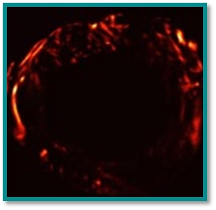
Intra myocardial vasculature can be mapped non-invasively in the beating heart, with a resolution of 300 µm in 2D using Ultra-fast Doppler imaging. This approach is highly dependent on image quality which varies from subject to subject and is limited to relatively large vessels. Ultrasonic Localization Microscopy (ULM) can map, for the very first time, blood vessels at the capillary scale (<50 µm) in depth. However, the ULM is very sensitive to movement. Both techniques are also limited to 2D imaging, which limits their diagnostic potential given the limited number of available echocardiographic windows.

TECHNOLOGY OVERVIEW

We propose a novel method to reconstruct super-resolved ultrasound angiograms in moving organs. The invention, the dynamic MULA, combines three technologies:
(a) Ultrasound Localization Microsocopy (ULM) for the tracking of highly echogenic, individual microbubbles (pre-injected) within the vasculature
(b) An ECG monitoring station is used to trigger intermittent ULM image acquisition, and more specifically the R-wave of the electrocardiogram.
(c) A mathematic model generates a Lagrangian beamformer based on non-rigid motion registration algorithm to form images directly in the myocardium’s material coordinates and thus correcting for the large myocardial motion and deformation.
The MULA framework enables the non-invasive imaging of the angioarchitecture and dynamics of intramyocardial flow in vessels as small as a few tens of microns in the rat’s beating heart in vivo. To our knowledge, this new method would be a first opportunity to assess these measurements directly, non-invasively and without ionization.
Preliminary results from a first clinical study will be available by the end of 2022.


COMPETITIVE ADVANTAGES

  • Non-invasive and no ionizing.
  • Mapping of the micro-vascularization (< 50 µm) of a moving organ.
  • Monitoring of perfusion dynamics during the cardiac cycle (functional data)
  • t=The system can be taken to the patient’s bedside


BUSINESS OPPORTUNITY

  • Technology available for in-licensing
  • Seeking for industrial partner for co-development
  • Eligibility to government financing for industry/academic maturation program


IP PROTECTION

CONTACTS

Jean Provost

PRINCIPAL INVESTIGATOR
Professor
Department of Engineering Physics
Polytechnique Montreal

Sébastien Bergeron, Ph. D.

CONTACT PERSON
Project Manager, Life Sciences
Axelys
sebastien.bergeron@axelys.ca