UNMET NEED
In patients with known or suspected coronary heart disease, cardiac imaging tests are often the first step in diagnosis and treatment planning. However, they remain for the most part limited to the coronary vasculature located on the surface of the heart, not to the intramyocardial vasculature located inside the myocardial muscle.
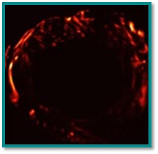
Intra myocardial vasculature can be mapped non-invasively in the beating heart, with a resolution of 300 µm in 2D using Ultra-fast Doppler imaging. This approach is highly dependent on image quality which varies from subject to subject and is limited to relatively large vessels. Ultrasonic Localization Microscopy (ULM) can map, for the very first time, blood vessels at the capillary scale (<50 µm) in depth. However, the ULM is very sensitive to movement. Both techniques are also limited to 2D imaging, which limits their diagnostic potential given the limited number of available echocardiographic windows.